We study how forces and nutrients around a tumor change the way cancer cells use energy and build protective structures. Our goal is to find weaknesses that help the immune system recognize and kill them.
We study the processes that help cancer cells survive and look for ways to disrupt them.
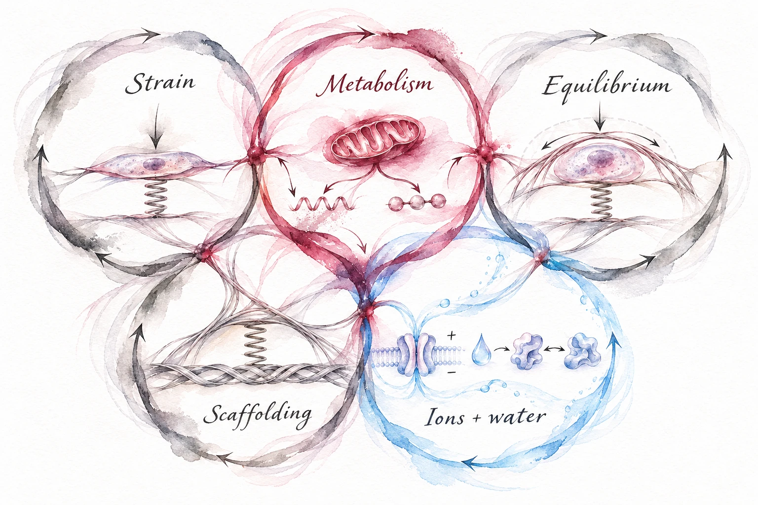
A CONNECTED VIEW OF THE CELL
Cells respond to their surroundings and change them in return.
A cell’s surroundings affect how it uses nutrients and builds materials. Those materials then change the conditions experienced by the cell and its neighbors.
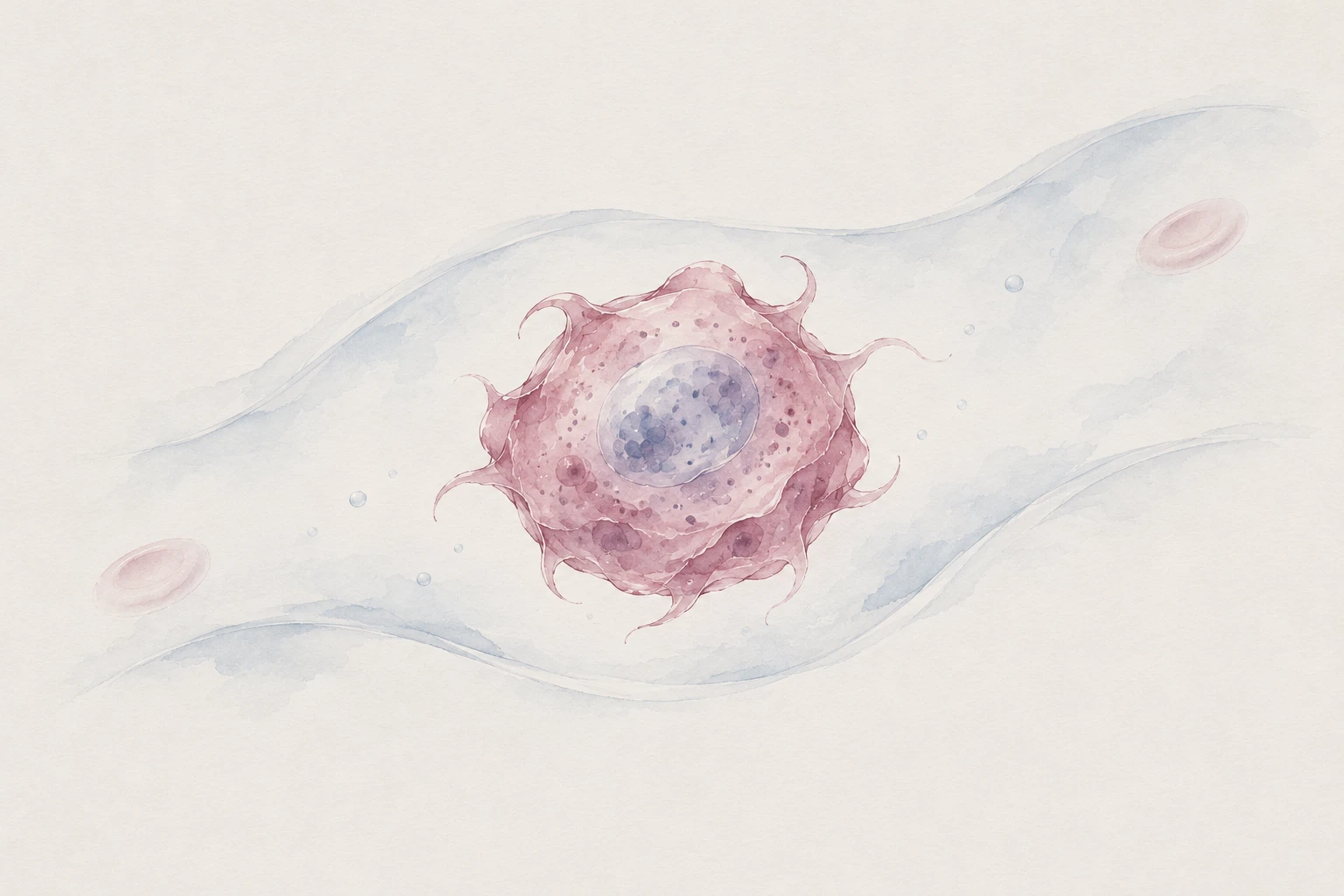
A conceptual illustration of the relationships we investigate.
Metabolism
Cells convert nutrients into energy and the materials they need to live.
Scaffolding
Cells build and reshape structures that support their own shape and the surrounding tissue.
Physical context
Cells experience forces and exchange water and ions with their surroundings.
FEATURED DISCOVERY · SCIENCE ADVANCES · 2026
A sugar-rich coat can help cancer cells resist immune attack.
What we found
We found that tissue stiffness and nutrient supply can change the sugar-rich coating on a cell’s surface, called the glycocalyx. In laboratory conditions designed to resemble the body, a stress-response protein called HSF1 helps cells use glucose to expand this coat.
Why it matters
In experimental models, interfering with this process made cancer cells easier for natural killer cells—a type of immune cell—to kill. This suggests a possible way to weaken cancer’s protection that needs further testing.
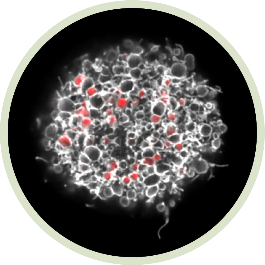
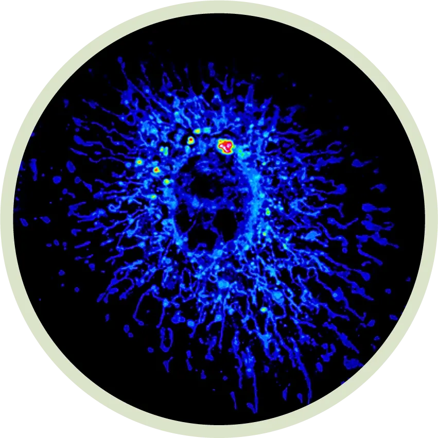
A single cell viewed through a microscope in the Tharp laboratory. Image from the Tharp laboratory; shown for context, not as a figure from this study.
FOLLOW THE CELL
What helps cancer cells survive as they spread through the body?
When cancer spreads from its original tumor to another organ, it is called metastasis. We study what cells need to survive each stage—and whether those needs reveal ways to stop them.
PUBLISHED FINDING
Cancer cells can build a sugar-rich coat that protects them from immune attack.
Inside the tumor
Cells respond to the stiffness of nearby tissue and the nutrients available to them.
How cells respond
These conditions can change how cells use nutrients to build their surface coat, called the glycocalyx.
Why this matters
In laboratory models, interfering with this process can make cancer cells easier for immune cells to kill.
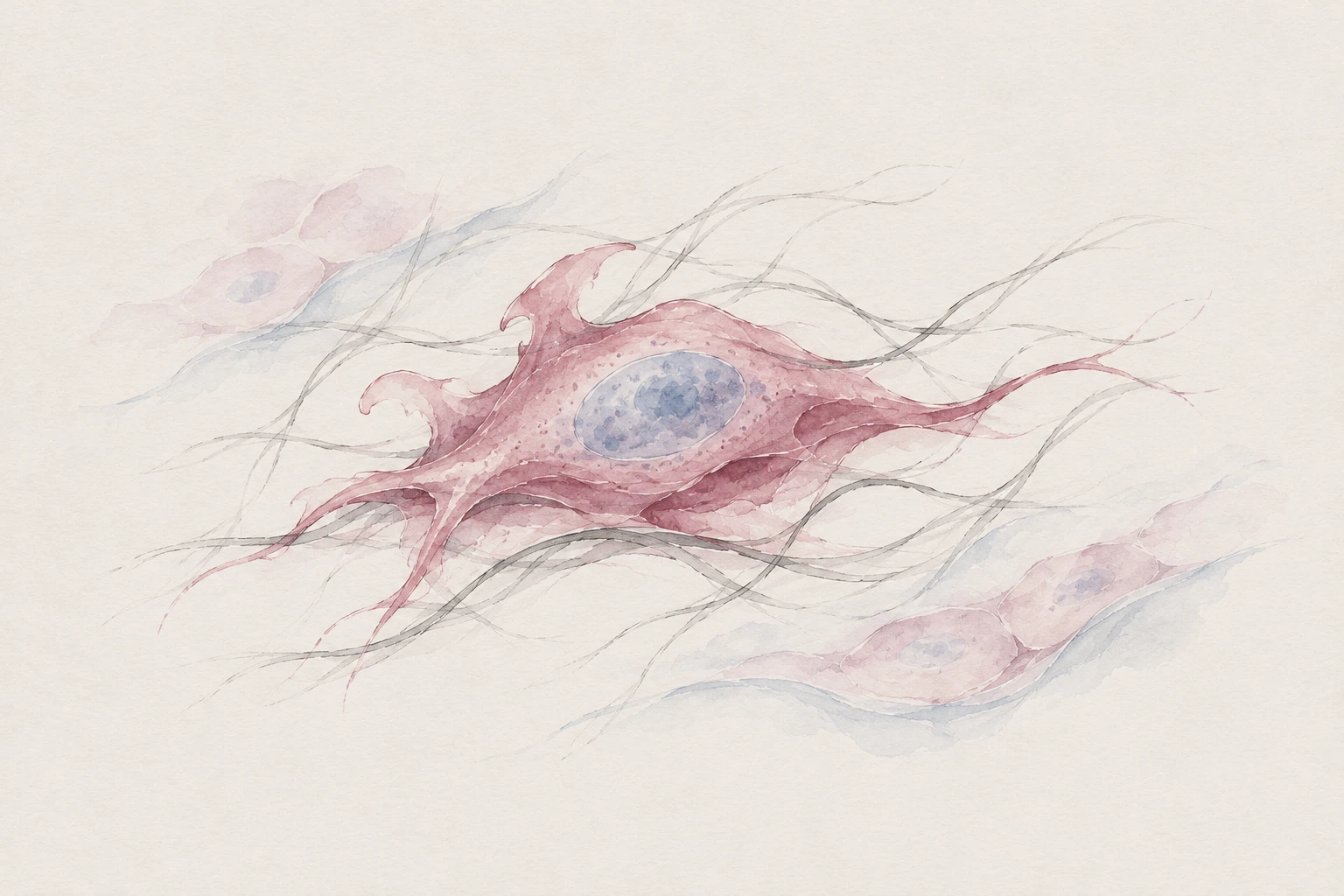
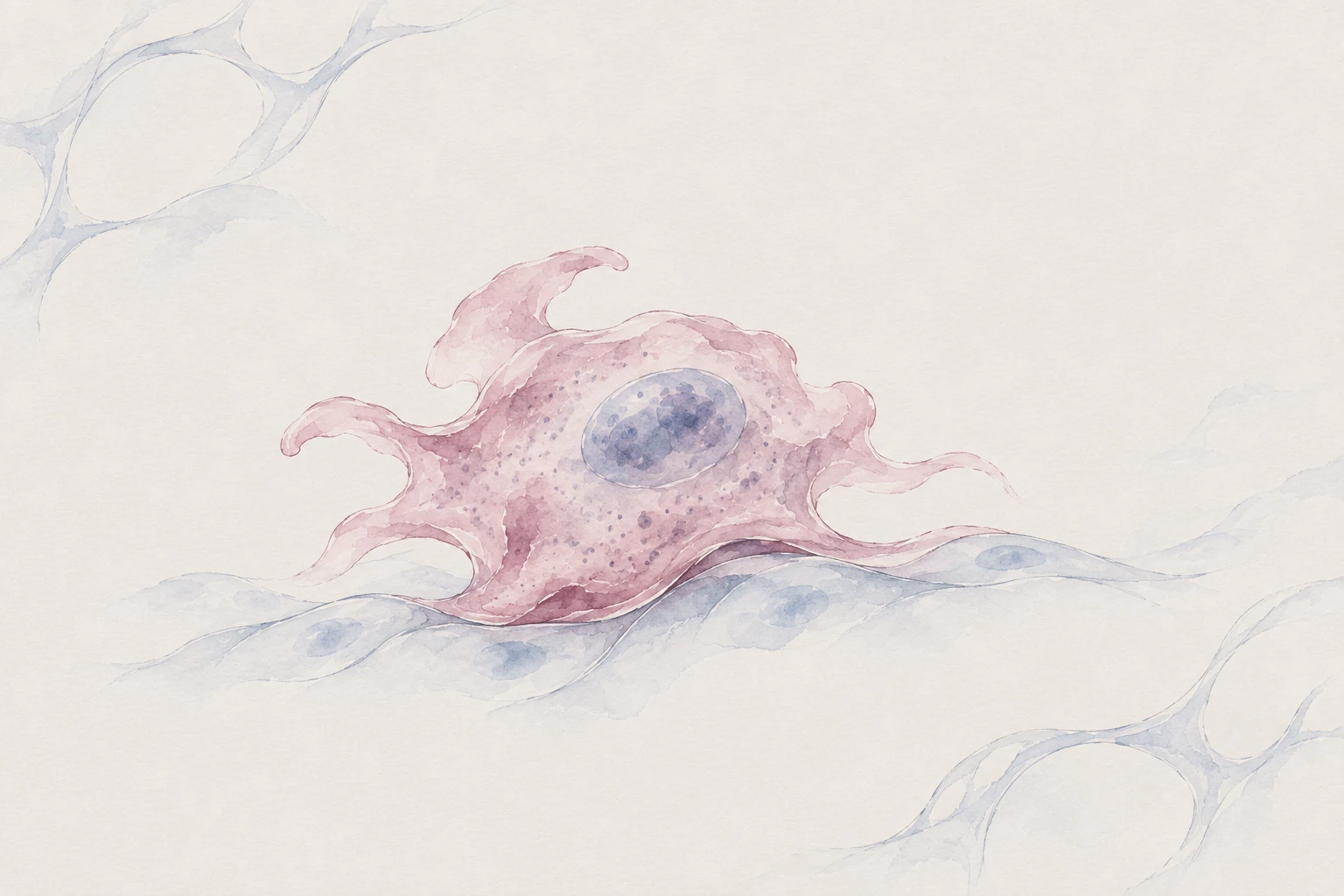
The watercolors are conceptual illustrations of our research questions.
THE WAY WE THINK
Cells change how they behave in response to their surroundings.
Cells detect how stiff their surroundings are and which nutrients are available. We ask how these signals change the way cells grow, move, and protect themselves.
Our work connects the forces cells experience, the ways they use nutrients, and the materials they build. We focus on two barriers that can protect tumors from immune cells: dense networks of collagen, a structural protein in tissue, and a sugar-rich coating on cancer cells called the glycocalyx.
By finding out what cells need to build and maintain these barriers, we aim to identify ways to weaken them and improve the immune system’s ability to attack cancer.
01 / RESEARCH
Three questions guide our cancer research.
Open a question to explore the projects behind it. Each story connects a biological problem to an opportunity to intervene.
01
What helps cancer cells spread to other parts of the body?
We study how cancer cells obtain the energy and materials they need to survive in different parts of the body.2 project stories
01 / MITOCHONDRIAL ADAPTABILITY
What’s under the hood of a metastatic cell?
A Toyota Camry and a Lamborghini Aventador have engines that operate on the same basic principle, yet deliver dramatically different performance on the track. Their components, configuration, and tuning determine what they can do—and what they require to do it.
For a metastatic cell, success depends on performing across remarkably different conditions: escaping a tumor, surviving transit, and establishing itself in unfamiliar tissue. We investigate how the composition and organization of mitochondrial machinery make this adaptability possible. By reverse engineering what equips these cells to succeed, we aim to identify—and disrupt—the metabolic requirements of their journey. Our goal is metastasis prophylaxis: preventing cancer cells from acquiring or sustaining the capabilities they need to spread.
How does cancer turn treatment stress into a survival strategy?
Stopping a production line can force a factory to reroute the work that keeps it running. We ask whether cancer cells respond to treatment in a similar way, redirecting metabolism to withstand stress and sustain essential cellular structures. In pancreatic cancer, we investigate how signals from the surrounding matrix influence these adaptations, including the production of nucleotides and sugar-based materials. By tracing where nutrients go as cells respond to treatment, we aim to identify the dependencies of surviving cells and block the adaptations that allow disease to persist.
02
How do tumors build barriers that protect them from immune cells?
We investigate how protective cell coatings and dense tissue can prevent immune cells from reaching or killing cancer cells.3 project stories
03 / GLYCO-IMMUNE SURVEILLANCE
Can a cancer cell build its own camouflage?
Camouflage changes what an observer can detect. Cancer cells can alter what the immune system encounters by changing the sugar-rich coat on their surface. We investigate how tissue mechanics, nutrient availability, and mitochondrial stress shape the production of this coat—and how those changes affect immune recognition and killing. We also ask whether cells carry this protection with them when they leave a tumor. By identifying the metabolic requirements for building and maintaining these surfaces, we aim to make cancer cells more vulnerable to immune clearance.
Can an immune cell reach what it needs to make an anti-tumor response?
A key can fit a lock perfectly and still be useless if it cannot reach the keyhole. Immune recognition faces a similar problem: a cancer cell may display the right molecular targets while presenting a surface that makes them difficult to engage. We study how cell shape, membrane folds, and surface coatings influence these encounters. We are developing probes with adjustable molecular reach to map which parts of a tumor cell are accessible, alongside models that reproduce the physical and nutrient conditions of human tissues. Our goal is to understand what turns molecular recognition into productive immune contact.
06 / FIBROSIS AND IMMUNE ACCESS
Who builds the barriers that keep immune cells out?
A security team cannot protect a building if the corridors keep it from reaching the threat. In tumors, collagen-rich networks can redirect immune cells and alter their activity. We study collagen-synthesizing tumor-associated macrophages and ask how their matrix production shapes immune access and function. We also examine which collagen–immune-cell contacts impede T-cell movement or function. By separating the builders, the materials, and the interactions that create exclusion, we aim to identify ways to restore effective immune access.
How can more realistic laboratory models help us find better treatments?
We recreate the physical conditions and nutrient supplies of tissues to study how cells behave inside the body.2 project stories
02 / CELLULAR SENSING
How does a cell read a world it cannot see?
Cells cannot see, but they can touch and taste. They probe the resistance of their surroundings and sample the nutrients available to them, reading a kind of biochemical braille. We study how these signals work together to guide metabolism and determine whether cells pull on their environment or build materials that change it. We ask how aging and metabolic stress alter this exchange, and whether responses that initially help cells cope can become self-reinforcing sources of fibrosis. By recreating the physical and chemical conditions of human tissues, we aim to understand where this conversation breaks down—and how to restore it.
A building can remain standing while its supporting structures gradually lose the ability to share a load. We ask whether a similar process unfolds in aging breast tissue. The cells lining mammary ducts maintain an open interior while staying connected to their neighbors. We test whether changes in the sugar-rich coat facing that interior shift more of the mechanical burden onto cell–cell junctions, altering tissue organization and cell identity. By identifying how this balance changes with age, we aim to find ways to preserve the architecture that helps keep tissues healthy.
We recreate conditions inside the body to study cancer more accurately.
Cells grown on hard plastic with standard nutrient mixtures can behave differently from cells inside the body. We build laboratory models that better reproduce real tissues so we can understand cancer and test possible treatments under more relevant conditions.
We control how stiff a cell’s surroundings are.
We use engineered materials to recreate the resistance cells feel when they attach to tissue. Live imaging lets us watch how changing these conditions changes cell behavior.
We match the nutrients cells encounter in the body.
Nutrient mixtures designed to resemble those in the body help us study responses that standard culture conditions can miss. We measure how cells use nutrients and which molecules they produce.
We study how tumor and immune cells affect one another.
We bring cancer cells, immune cells, and cells that support tissues into shared experimental systems. We also use mouse models to study how these interactions affect tumors.
Our institute helped explain why cell attachment matters.
Our lab joined an institute that helped establish how a cell’s connection to its surroundings governs its survival.
Founded by William and Lillian Fishman in 1976 as the La Jolla Cancer Research Foundation, today’s Sanford Burnham Prebys became a home for foundational discoveries in cell adhesion and cancer biology.
Kevin established the Tharp Laboratory here in November 2023. Our work carries this scientific tradition into questions of metabolic adaptation, metastasis, and immune recognition.
Cells use specific molecules to attach to their surroundings
Michael Pierschbacher and Erkki Ruoslahti identified the RGD cell-recognition sequence in fibronectin. With Robert Pytela, they then isolated a fibronectin receptor—work that helped establish integrin-mediated binding to the extracellular matrix.
Steven Frisch and Hunter Francis described anoikis: programmed cell death triggered when normal epithelial cells lose contact with their matrix. Their study linked escape from this response to the acquisition of anchorage independence.
Frisch, Kristiina Vuori, Ruoslahti, and P. Y. Chan-Hui showed that activated focal adhesion kinase can suppress anoikis, connecting integrin-associated signaling to cell survival.
We ask how changing physical and nutritional environments reshape metabolism—and how those adaptations help cancer cells survive, spread, and evade immunity.
Our team brings different skills to shared scientific questions.
Our team combines expertise in how cells use nutrients, how tissues are built, and how the immune system responds to cancer.
PRINCIPAL INVESTIGATOR
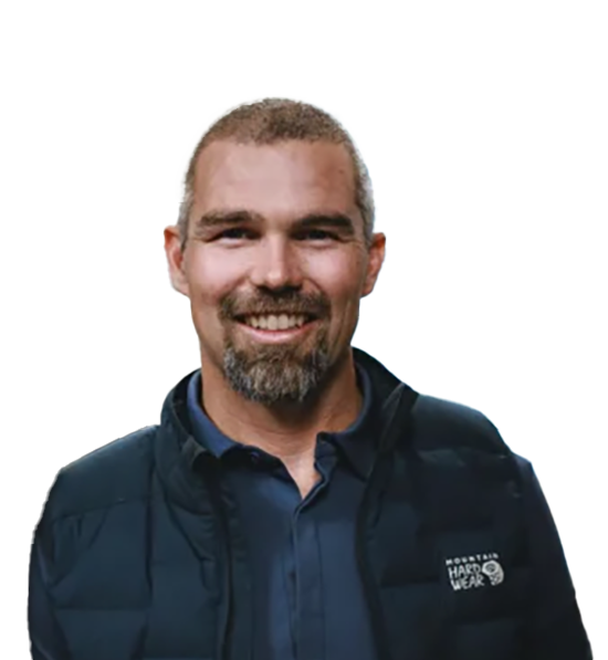
Kevin M. Tharp, PhD
Assistant Professor Cancer Metabolism and Microenvironment Program Sanford Burnham Prebys
Kevin studies how cells use metabolic programs to adapt to their physical surroundings. His research connects mitochondrial biology, tissue mechanics, and immune evasion to identify opportunities to interfere with cancer progression.
At UC Berkeley, Kevin worked with Andreas Stahl and Kevin Healy on biomaterials, stem cell differentiation, and thermogenic adipose tissues. He then joined Valerie Weaver at UCSF to study how cellular and tissue mechanics affect mitochondrial metabolism. He established the Tharp Laboratory in November 2023.